A 65 year-old man with atrial fibrillation developed the acute onset of left hemiplegia and a right preference. Initial MRI diffusion scan indicated a large right MCA infarct. Two days later, the patient became lethargic.
![]()
![]()
![]()
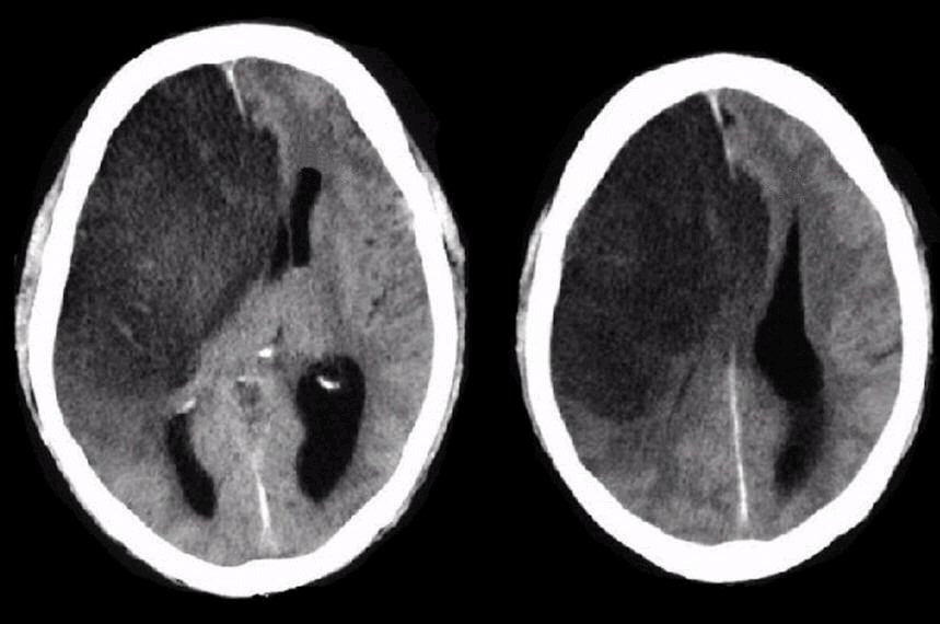
Axial CT scan: Note the large well defined infarction in the distribution of the right proximal MCA. However, also note the herniation of the right cingulate gyrus under the falx then compressing the left frontal lobe. Among other findings, this is an ominous sign denoting significant mass effect and increased intracranial pressure. Also note the amount of midline shift of the right hemisphere on the left hemisphere. One using way of measuring midline shift is looking at how much the pineal gland has shifted off the midline. The pineal gland is normally in the midline, and in most adults, it will calcify, allowing it to be easily seen on CT imaging.
Revised
04/20/06.
The Electronic Curriculum is copyrighted 1998, Case Western Reserve University
School of Medicine.